Home
/ Hip And Upper Thigh Anatomy - The anatomy of the legs | Leg anatomy, Muscle anatomy ... - Aug 28, 2020 · this is the only quad muscle with two heads, and that crosses two joints — the hip and the knee — to assist with both knee extension and hip flexion.
Hip And Upper Thigh Anatomy - The anatomy of the legs | Leg anatomy, Muscle anatomy ... - Aug 28, 2020 · this is the only quad muscle with two heads, and that crosses two joints — the hip and the knee — to assist with both knee extension and hip flexion.
Hip And Upper Thigh Anatomy - The anatomy of the legs | Leg anatomy, Muscle anatomy ... - Aug 28, 2020 · this is the only quad muscle with two heads, and that crosses two joints — the hip and the knee — to assist with both knee extension and hip flexion.. The iliopsoas muscle, which extends from the lower back to upper femur; The adductor muscle on the inner thigh; Aug 28, 2020 · this is the only quad muscle with two heads, and that crosses two joints — the hip and the knee — to assist with both knee extension and hip flexion. In human anatomy, the thigh is the area between the hip and the knee.anatomically, it is part of the lower limb. Jul 06, 2021 · hip joint:
Stabilizes head of femur in acetabulum quadratus femoris quadratus femoris is a flat and rectangular shaped muscle that is the most inferior gluteal muscle of the deep layer, lying inferior to the gemellus inferior. In human anatomy, the thigh is the area between the hip and the knee.anatomically, it is part of the lower limb. Hip, & thigh 2021 knee & distal lower limb 2021. Jul 06, 2021 · hip joint: In this section, learn more about the upper limb:
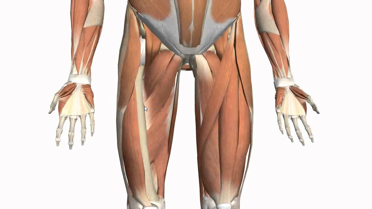
Muscles of the Thigh and Gluteal Region - Part 2 - Anatomy ... from i.ytimg.com Hip, & thigh 2021 knee & distal lower limb 2021. The hand is a very mobile part of the upper limb, and we perform very specialised tasks with it every day, key adaptations can be seen in the specialised structures of the hand. Originating at two different points on your pelvis, the rectus femoris passes down the front of your thigh and ends at its attachment point on the kneecap (patella). Hip muscles the hip joint is surrounded by several muscles, including: Stabilizes head of femur in acetabulum quadratus femoris quadratus femoris is a flat and rectangular shaped muscle that is the most inferior gluteal muscle of the deep layer, lying inferior to the gemellus inferior. Its bones, muscles, nerves, joints, blood vessels and lymphatics, anatomical areas, and structures found in the hand. Jun 28, 2021 · hip joint (overview) the hip joint is a large ball and socket synovial joint between the head of the femur and the acetabulum of the pelvis.it is structured in such a way that enables movement in all axes, while bearing both our summer and winter body mass and providing stability for the body during movement. Aug 28, 2020 · this is the only quad muscle with two heads, and that crosses two joints — the hip and the knee — to assist with both knee extension and hip flexion.
Stabilizes head of femur in acetabulum quadratus femoris quadratus femoris is a flat and rectangular shaped muscle that is the most inferior gluteal muscle of the deep layer, lying inferior to the gemellus inferior.
Thigh external rotation, thigh abduction (from flexed hip), thigh adduction (secondary function); In human anatomy, the thigh is the area between the hip and the knee.anatomically, it is part of the lower limb. The anatomy of the shoulder girdle consists of several joints, or articulations, which connect the upper limb to the rest of the skeleton.you may also see this referred to as the pectoral girdle in some textbooks. The hand is a very mobile part of the upper limb, and we perform very specialised tasks with it every day, key adaptations can be seen in the specialised structures of the hand. Anatomy of the hip an inside look at the structure of the hip. Hip muscles the hip joint is surrounded by several muscles, including: Quadriceps, a group of four muscles that comprise the front of the thigh; In this section, learn more about the upper limb: Hip, & thigh 2021 knee & distal lower limb 2021. Stabilizes head of femur in acetabulum quadratus femoris quadratus femoris is a flat and rectangular shaped muscle that is the most inferior gluteal muscle of the deep layer, lying inferior to the gemellus inferior. Jul 06, 2021 · hip joint: Its bones, muscles, nerves, joints, blood vessels and lymphatics, anatomical areas, and structures found in the hand. The iliopsoas muscle, which extends from the lower back to upper femur;
Jul 06, 2021 · hip joint: The hip bones have three main articulations: The anatomy of the shoulder girdle consists of several joints, or articulations, which connect the upper limb to the rest of the skeleton.you may also see this referred to as the pectoral girdle in some textbooks. Thigh external rotation, thigh abduction (from flexed hip), thigh adduction (secondary function); In human anatomy, the thigh is the area between the hip and the knee.anatomically, it is part of the lower limb.
Muscles In Lower Back And Hip / Daily Health Post: 6 ... from www.anatomyfacts.com The adductor muscle on the inner thigh; Gluteal muscles, located on the back of the hip (buttocks); Jun 28, 2021 · hip joint (overview) the hip joint is a large ball and socket synovial joint between the head of the femur and the acetabulum of the pelvis.it is structured in such a way that enables movement in all axes, while bearing both our summer and winter body mass and providing stability for the body during movement. The single bone in the thigh is called the femur.this bone is very thick and strong (due to the high proportion of bone tissue), and forms a ball and socket joint at the hip, and a modified hinge joint at the knee. Aug 28, 2020 · this is the only quad muscle with two heads, and that crosses two joints — the hip and the knee — to assist with both knee extension and hip flexion. Anatomy of the hip an inside look at the structure of the hip. Hip, & thigh 2021 knee & distal lower limb 2021. Thigh external rotation, thigh abduction (from flexed hip), thigh adduction (secondary function);
In human anatomy, the thigh is the area between the hip and the knee.anatomically, it is part of the lower limb.
Jun 28, 2021 · hip joint (overview) the hip joint is a large ball and socket synovial joint between the head of the femur and the acetabulum of the pelvis.it is structured in such a way that enables movement in all axes, while bearing both our summer and winter body mass and providing stability for the body during movement. In this section, learn more about the upper limb: Originating at two different points on your pelvis, the rectus femoris passes down the front of your thigh and ends at its attachment point on the kneecap (patella). The iliopsoas muscle, which extends from the lower back to upper femur; Gluteal muscles, located on the back of the hip (buttocks); The single bone in the thigh is called the femur.this bone is very thick and strong (due to the high proportion of bone tissue), and forms a ball and socket joint at the hip, and a modified hinge joint at the knee. Jul 06, 2021 · hip joint: The anatomy of the shoulder girdle consists of several joints, or articulations, which connect the upper limb to the rest of the skeleton.you may also see this referred to as the pectoral girdle in some textbooks. Its bones, muscles, nerves, joints, blood vessels and lymphatics, anatomical areas, and structures found in the hand. Hip, & thigh 2021 knee & distal lower limb 2021. Anatomy of the hip an inside look at the structure of the hip. Thigh external rotation, thigh abduction (from flexed hip), thigh adduction (secondary function); The adductor muscle on the inner thigh;
Jul 06, 2021 · hip joint: Quadriceps, a group of four muscles that comprise the front of the thigh; The hand is a very mobile part of the upper limb, and we perform very specialised tasks with it every day, key adaptations can be seen in the specialised structures of the hand. Anatomy of the hip an inside look at the structure of the hip. Originating at two different points on your pelvis, the rectus femoris passes down the front of your thigh and ends at its attachment point on the kneecap (patella).
Human Anatomy and Physiology of Muscles Online on | Human ... from s-media-cache-ak0.pinimg.com Quadriceps, a group of four muscles that comprise the front of the thigh; Jun 28, 2021 · hip joint (overview) the hip joint is a large ball and socket synovial joint between the head of the femur and the acetabulum of the pelvis.it is structured in such a way that enables movement in all axes, while bearing both our summer and winter body mass and providing stability for the body during movement. Anatomy of the hip an inside look at the structure of the hip. Aug 28, 2020 · this is the only quad muscle with two heads, and that crosses two joints — the hip and the knee — to assist with both knee extension and hip flexion. Originating at two different points on your pelvis, the rectus femoris passes down the front of your thigh and ends at its attachment point on the kneecap (patella). In this section, learn more about the upper limb: The adductor muscle on the inner thigh; The single bone in the thigh is called the femur.this bone is very thick and strong (due to the high proportion of bone tissue), and forms a ball and socket joint at the hip, and a modified hinge joint at the knee.
The single bone in the thigh is called the femur.this bone is very thick and strong (due to the high proportion of bone tissue), and forms a ball and socket joint at the hip, and a modified hinge joint at the knee.
The hip bones have three main articulations: Hip, & thigh 2021 knee & distal lower limb 2021. The anatomy of the shoulder girdle consists of several joints, or articulations, which connect the upper limb to the rest of the skeleton.you may also see this referred to as the pectoral girdle in some textbooks. Stabilizes head of femur in acetabulum quadratus femoris quadratus femoris is a flat and rectangular shaped muscle that is the most inferior gluteal muscle of the deep layer, lying inferior to the gemellus inferior. Aug 28, 2020 · this is the only quad muscle with two heads, and that crosses two joints — the hip and the knee — to assist with both knee extension and hip flexion. Jun 28, 2021 · hip joint (overview) the hip joint is a large ball and socket synovial joint between the head of the femur and the acetabulum of the pelvis.it is structured in such a way that enables movement in all axes, while bearing both our summer and winter body mass and providing stability for the body during movement. Originating at two different points on your pelvis, the rectus femoris passes down the front of your thigh and ends at its attachment point on the kneecap (patella). The hand is a very mobile part of the upper limb, and we perform very specialised tasks with it every day, key adaptations can be seen in the specialised structures of the hand. Thigh external rotation, thigh abduction (from flexed hip), thigh adduction (secondary function); In this section, learn more about the upper limb: Anatomy of the hip an inside look at the structure of the hip. Hip muscles the hip joint is surrounded by several muscles, including: The adductor muscle on the inner thigh;
Stabilizes head of femur in acetabulum quadratus femoris quadratus femoris is a flat and rectangular shaped muscle that is the most inferior gluteal muscle of the deep layer, lying inferior to the gemellus inferior upper thigh anatomy. Quadriceps, a group of four muscles that comprise the front of the thigh;